Past, present & future of healthcare services in India

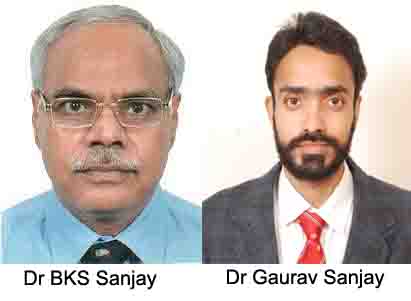
Monday, 12 June 2023 | Dr BKS Sanjay & Dr Gaurav Sanjay | in Guest Column
GUEST COLUMN

Being universal and inevitable, change has also been seen in the healthcare services in India. The senior author had entered medical college at Kanpur in 1974 while the second author joined medical college at Dehradun in 2003.
At the first author’s medical college the lecture halls were huge and could accommodate nearly 400 students, usually two batches of about 200 students each. The back benchers could hardly understand the words written on the blackboard and could barely hear the spoken words most of the time, particularly if the teacher was soft spoken with a low tone of voice. But nowadays the methodology of teaching has changed drastically not only in the higher education institutes but also in the primary schools.
Similarly, the clinical teaching five decades ago was mainly patient based. It was difficult to even see the involved part of the body and learning clinical examination was a distant dream in front of the professor. However, there was a well-established tradition of interactive evening teaching in the wards by the PG students, usually demonstrators and registrars. Ward round-based teaching was a good, convenient and beneficial method of teaching because it was an interactive method-based teaching. There wasn’t a huge age difference among the postgraduates/demonstrators and the MBBS students, therefore, they were very friendly and helpful. Students were not hesitant in clearing their doubts and asking questions. We personally feel this kind of interactive clinical teaching should be continued and should be started if it’s not being done. In clinical practice there was a lot of physical work in those days which was required by junior residents or PG students like collection of blood samples, arranging other investigations, blood donors and blood units, part preparation of the surgical site, cleaning and painting of the part before the surgery, assisting the surgeon by just holding the re-tractors, application and removal of the plaster cast, transporting the patient between recovery and OT which were a part of the duty of the residents. Now, things have changed drastically because of computerisation and automation of the laboratories and more investment in healthcare professionals such as phlebotomist, ward attendants, various technicians and paramedics etc and other specialised healthcare services.
Being orthopaedic surgeons, we would like to mention a few things from a surgeon’s perspective. Though the hospital wards and operation theatres were cleaner than our homes, they were never sterile because there was no concept of sterilisation of OT air. The usual protocol was to clean the operating table and to autoclave the gloves, syringes, needles, OT drapes, linen and the operating instruments. When the senior author started his MS (Orthopaedic) training in PGI Chandigarh in 1981, he saw for the first time that the operating theatres were being fumigated every day in the night before the start of any surgery. Even a premier institute like PGI Chandigarh was lacking the facility of fluoroscopy or image intensifier in operation theatre. Although fracture of neck femur was not a very common surgery it needed an image intensifier or X- ray control to check the position of fracture reduction and the position of screws within the bone. The only facility we had at that time was a mobile X-ray machine in the operation theatre complex which was being shared among many surgical specialties which could be requested on call. The technician from the X-ray department would take the radiographic exposure in the operation theatre after shifting the heavy machine and return to the department’s darkroom to develop the film manually. The whole process used to take a minimum of 20-30 minutes. During that time, the surgery would be put on hold, which may increase the risk of bleeding and infection. Currently almost all orthopaedic operation theatres have an image intensifier which can give the X-ray image with just a click. Images can also be stored permanently and can be retrieved instantly as it is all computerised.
Advancement in fracture treatment is another aspect we will like to mention here. Fifty years ago all difficult fractures were reduced after opening the skin and then fixing it. The drilling of the bone was done manually which was usually very tiring and at times surgeons would reduce the number of screws ideally required for fixation due to surgeon fatigue. Most of the time the reduction and fixation were accepted even though it was not satisfactory or perfect. The availability of the preferred type, size and quality of the implants, electric and pneumatic (air) drill and saw, and image intensifier in operation theatre has improved the quality of fracture treatment. Even surgeons with average surgical skills are now able to give excellent results. In certain cases, it becomes difficult to say if the patient had any fracture earlier.
There has been tremendous improvement in cancer surgery. Initially in orthopaedic surgery, the usual treatment of bone and soft tissue tumors used to be amputations of affected limbs which caused permanent disabilities. The invention of CT, MRI, PET and bone scan has improved the quality of surgery, post-operative prognosis and quality of post cancer therapy survival. Nowadays the limb salvage surgery is a norm.
A 15-year-old girl was suffering from bone cancer of the leg bone and was treated 17 years ago by the authors in Dehradun. The bone tumor had been removed, the defect had been reconstructed by recycling the same bone and was used as bone graft and reconstruction with a nail was done. This patient was treated post surgery with chemotherapy in PGI, Chandigarh. After 17 years of the surgery, she has a near normal personal and social life while working as a teacher. In her case the credit goes to the quality of surgery, chemotherapy and faith of the family in the doctors. This is not an isolated case but many limbs and lives are being saved nowadays due to technological and surgical advancement.
Total joint replacement surgery is another major development. Fifty years ago a patient with any kind of arthritis had to rely mainly on painkillers. When the first author was in Kanpur Medical College he never saw any surgery for arthritis but PGI Chandigarh was one of the few centres in the country where joint replacement surgery was started in 1981. The hip and knee joint replacement surgeries changed the lives of not only the patients but the professional lives of orthopaedic surgeons too. Similarly, spine surgery which was a rare and risky surgery is nowadays a common and simple surgery which gives gratifying results. When the first author joined orthopaedic surgery, it was not a very popular branch. The most sought speciality used to be internal medicine and paediatrics but three decades later when the second author joined his PG training, orthopaedic surgery was one of the most desired specialities among male doctors. It is just behind radiology which was the least preferred speciality for post-graduation 50 years ago.
The major developments which have occurred in radiology, especially in imaging- CT, MRI, PET, bone scan, interventional radiology and availability of the various isotopes etc have occurred in the last 50 years. Minimally invasive surgery, interventional surgery, navigation and robotic surgery have made tremendous contributions in both medical and surgical fields. CAD (computer aided design) software has facilitated the manufacturing of implants. Nowadays, 3D printed models give a better understanding of complex pathology and anatomy of the patient which helps to do reconstruction and plan the surgeries with patient tailored implants for perfect results. 3D printed patient specific models in cardiothoracic surgery, kidney and other organs transplantation including bone transplantation are the newer advancement in healthcare services. Artificial intelligence (AI) in our opinion has the potential to change medical practice in the future.
Technology has made the life of doctors, technicians, engineers easier and it has improved the quality of life of many patients. The smart watch is a small and relatively cheap and simple tool to monitor the vital parameters of the body. In the earlier days one had to go to a clinic to know the current BP, temperature, blood sugar etc but nowadays a smart watch or certain portable devices can give you 24 hours continuous monitoring of the vital parameters which can help in prevention of serious complications of the various health issues.
(The authors are orthopaedic surgeons based in Dehradun. Views expressed are personal)


